Image of Mcvaughia
Description:
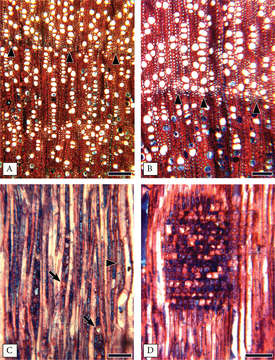
Figure 1. Wood anatomy of Mcvaughiasergipana. A–B transverse sections: Growth rings marked by radially narrow fibers (arrowheads) and a discontinuous line of axial parenchyma (in B); vessels are narrow and abundant, arranged in radial rows of 4 or more cells; some solitary vessels present; parenchyma rare, paratracheal scanty or at the growth ring limits; heartwood vessels in the bottom with content C radial section: Rays 2–3 cells wide, non-storied; prismatic crystals present in ray cells (arrows); parenchyma with 3 cells per strand (arrowhead) D ray heterocellular with procumbent, square and upright cells mixed throughout the ray. Scale bars: 150 μm (A), 100 μm (B–C), 60 μm (D).
Included On The Following Pages:
- Life (creatures)
- Cellular (cellular organisms)
- Eukaryota (eukaryotes)
- Archaeplastida (plants)
- Chloroplastida (green plants)
- Spermatophytes (seed plants)
- Angiosperms (Dicotyledons)
- Eudicots
- Superrosids
- Rosids
- Malpighiales
- Malpighiaceae (Barbados cherry family )
- Mcvaughia
- NO NAME!
This image is not featured in any collections.
Source Information
- license
- cc-by-3.0
- copyright
- Rafael F. Almeida, Isabel R. Guesdon, Marcelo R. Pace, Renata M.S. Meira
- bibliographic citation
- Almeida R, Guesdon I, Pace M, Meira R (2019) Taxonomic revision of Mcvaughia W.R.Anderson (Malpighiaceae): notes on vegetative and reproductive anatomy and the description of a new species PhytoKeys (117): 45–72
- original
- original media file
- visit source
- partner site
- Phytokeys
- ID