portrait
Description:
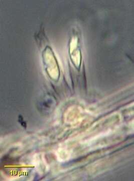
Portrait of colonial form of chrysophyte flagellate, Epipyxis. Cells attach to the base of vase-like loricae by protoplasmic threads containing microtubules. Loricae are constructed of overlapping scales. The scales, visible only by electron-microscopy or staining are composed of interwoven microfibrils. Yellow chloroplast with small stigma (not well-seen in this image). Epipyxis is mixotrophic. Phagotrophy involves bacterial capture by the longer of the two flagella and formation of a feeding "basket" by microtubular action at the anterior of the cell. Often epiphytic on filamentous algae as seen here but sometimes free-swimming. From freshwater pond near Boise, Idaho. Phase contrast.
Included On The Following Pages:
- Life (creatures)
- Cellular (cellular organisms)
- Eukaryota (eukaryotes)
- SAR (Stramenopiles, Alveolates, Rhizaria)
- Stramenopiles (heterokont)
- Ochrophyta (Ochrophyte)
- Chrysophyceae (golden algae)
- Chromulinales
- Dinobryaceae
- Epipyxis
- Oomycota (oomycetes)
- Chrysista
This image is not featured in any collections.
Source Information
- license
- cc-by-nc
- author
- William Bourland
- provider
- micro*scope
- original
- original media file
- visit source
- partner site
- micro*scope
- ID