whole mount
Description:
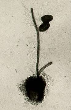
Single cell, fixed with osmikum vapor and imaged using a transmission electron microscope. The cell is coated in small scales. There are two flagella, the longer one is coated with (tripartite) hairs that help draw food (bacteria, two of which lie beside the flagellum) into the cell.
Included On The Following Pages:
- Life (creatures)
- Cellular (cellular organisms)
- Eukaryota (eukaryotes)
- SAR (Stramenopiles, Alveolates, Rhizaria)
- Stramenopiles (heterokont)
- Ochrophyta (Ochrophyte)
- Chrysophyceae (golden algae)
- Chromulinales
- Paraphysomonadaceae
- Paraphysomonas
- Paraphysomonas butcheri
- Oomycota (oomycetes)
- Chrysista
This image is not featured in any collections.
Source Information
- license
- cc-by-nc
- author
- D. J. Patterson.
- provider
- micro*scope
- original
- original media file
- visit source
- partner site
- micro*scope
- ID