eyespot
Description:
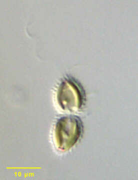
Portrait of Chrysodidymus synuroideus, a colonial synurophyte chrysophyte. Chrysodidymus is a monospecific genus. Two cells united at their bases form the colonies. The cells are conical. Each is covered by siliceous scales. The scales are formed in the chloroplast endoplasmic reticulum and transported to the cell surface. There are two flagella, one long (bearing tripartite hairs typical of stramenopiles on electron microscopy) and one short. There are two golden-colored parietal chloroplasts without pyrenoids. A posterior storage vacuole may contain chrysolaminarin. Red pigment granules can be seen at the apices of these cells. Swimming movement is a distinctive back and forth motion. From a freshwater irrigation ditch near McCall, Idaho. Differential interference contrast.
Included On The Following Pages:
- Life (creatures)
- Cellular (cellular organisms)
- Eukaryota (eukaryotes)
- SAR (Stramenopiles, Alveolates, Rhizaria)
- Stramenopiles (heterokont)
- Ochrophyta (Ochrophyte)
- Synurales (synurids)
- Mallomonadaceae
- Synura
- Synura synuroidea
- Oomycota (oomycetes)
- Chrysista
- Chrysophyceae (golden algae)
This image is not featured in any collections.
Source Information
- license
- cc-by-nc
- author
- William Bourland
- provider
- micro*scope
- original
- original media file
- visit source
- partner site
- micro*scope
- ID