portrait
Description:
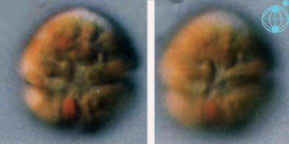
Durinskia (dew-rink-see-a) baltica (Levander 1894) Carty and Cox 1986. The images show cells in ventral view. The red stigma is visible in the sulcal area. The plastids are yellow-brown and multiple. The cingulum is in the middle of the cell.
Included On The Following Pages:
- Dinoflagellata (dinoflagellates)
- Life (creatures)
- Cellular (cellular organisms)
- Eukaryota (eukaryotes)
- SAR (Stramenopiles, Alveolates, Rhizaria)
- Alveolata (alveolates)
- Dinophyceae
- Peridiniales
- Kryptoperidiniaceae
- Durinskia
- Durinskia baltica
This image is not featured in any collections.
Source Information
- license
- cc-by-nc
- author
- Mona Hoppenrath and Shauna Murray
- provider
- micro*scope
- original
- original media file
- visit source
- partner site
- micro*scope
- ID