Silverline system
Description:
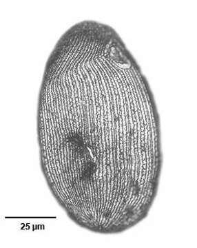
Right lateral view of the silverline system of the hymenostome ciliate Colpidium kleini (Foissner, 1969). C. kleini is very similar in overall appearance to C. colpoda although usually more slender and with fewer somatic kineties. The cytostome is in the anterior 1/4 of the cell. There is a curved paraoral membrane along the convex right margin of the cytostome. The left margin is slightly concave. There are three adoral membranelles. There are 32 to 44 somatic kineties. The kineties to the right and left of the oral aperture meet at a curved preoral suture.The right somatic kineties bend leftward at the level of the cytostome. There is an anterior apical area bare of cilia. There are rows of inconspicuous mucocysts between the somatic kineties. The ellipsoid macronucleus and adjacent micronucleus are centrally located. The single contractile vacuole is located in the midbody with a single excretory pore on the right surface. The feature most clearly distinguishing Colpidium kleini from C. coploda is the silverline system. In C. kleini there is only one secondary meridian (silverline) between two primary meridians (primary meridians correspond to somatic kineties). In some cases short segments of the secondary meridians may be duplicated. Short transverse L or T-shaped branches arise from both primary and secondary meridians at irregular intervals.Stained by the dry silver nitrate technic (see Foissner, W.Europ. J. Protistol.27,313-330;1991). Collected from an organically enriched freshwater pond near Boise, Idaho. Brightfield. Black and white.
Included On The Following Pages:
- Life (creatures)
- Cellular (cellular organisms)
- Eukaryota (eukaryotes)
- SAR (Stramenopiles, Alveolates, Rhizaria)
- Alveolata (alveolates)
- Ciliophora (ciliates)
- Intramacronucleata
- Oligohymenophorea
- Hymenostomatida
- Tetrahymenina
- Turaniellidae
- Colpidium
- Colpidium kleini
This image is not featured in any collections.
Source Information
- license
- cc-by-nc
- author
- William Bourland
- provider
- micro*scope
- original
- original media file
- visit source
- partner site
- micro*scope
- ID