Image of Coarazuphium whiteheadi Ball & Shpeley 2013
Description:
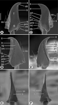
Figure 6.SEM micrographs of mandibles of Coarazuphium whiteheadi, new species. A and D dorsal aspect B and E ventral aspect C and F lateral aspect. Legend: art, anterior retinacular tooth; B, basal area; bb, basal brush; it, incisor tooth; mt, molar tooth; pmt, premolar tooth; prt, posterior retinacular ridge; rr, retinacular ridge; s, scrobe; ss, scrobal seta; str, supraterebral ridge; T, terebra; tr, terebral ridge; tt, terebral tooth; vg, ventral groove; vm, ventral microtrichia. Scale bars = 100 µm.
Included On The Following Pages:
- Life (creatures)
- Cellular (cellular organisms)
- Eukaryota (eukaryotes)
- Opisthokonta (opisthokonts)
- Metazoa (Animal)
- Bilateria
- Protostomia (protostomes)
- Ecdysozoa (ecdysozoans)
- Arthropoda (arthropods)
- Pancrustacea
- Hexapoda (hexapods)
- Insecta (insects)
- Pterygota (winged insects)
- Neoptera (neopteran)
- Endopterygota (endopterygotes)
- Coleoptera (beetles)
- Adephaga (adephagans)
- Carabidae (ground beetles)
- Coarazuphium
- Coarazuphium whiteheadi
- Panarthropoda
This image is not featured in any collections.
Source Information
- license
- cc-by-3.0
- copyright
- George E. Ball, Danny Shpeley
- bibliographic citation
- Ball G, Shpeley D (2013) Western Hemisphere Zuphiini: descriptions of Coarazuphium whiteheadi, new species, and Zuphioides, new genus, and classification of the genera (Coleoptera, Carabidae) ZooKeys 315: 17–54
- original
- original media file
- visit source
- partner site
- Zookeys
- ID