Image of Ferrocina Glover & J. D. Taylor 2007
Description:
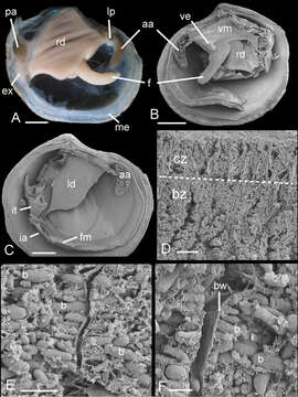
Figure 8.Ferrocina brunei sp. n. A Body from right side. Scale bar = 1 mm B Body from left side, mantle and left demibranch removed showing foot and visceral extension. Scale bar = 1 mm C Body from right side with visceral mass, right demibranch and mantle removed. Scale bar = 1 mm D Section through part of demibranch showing ciliated and bacteriocyte zones. Scale bar = 20 µm E Part of ctenidial filament in bacteriocyte zone showing bacteria. Scale bar = 5 µm F Detail of bacteriocytes and bacteria aligned normal to the apical cell wall. Scale bar = 2 µm. aa, anterior adductor muscle. b, bacteria. bw, bacteriocyte apical wall. bz, bacteriocyte zone. cz, ciliated zone. ex, exhalant aperture. f, foot. fm, fused mantle. ia, inhalant aperture. it, inverted tube of posterior exhalant aperture. ld, left demibranch. lp, labial palps. me, mantle edge. pa, posterior adductor muscle. rd, right demibranch. ve, visceral extension. vm, visceral mass.
Included On The Following Pages:
- Life (creatures)
- Cellular (cellular organisms)
- Eukaryota (eukaryotes)
- Opisthokonta (opisthokonts)
- Metazoa (Animal)
- Bilateria
- Protostomia (protostomes)
- Spiralia (spiralians)
- Mollusca (molluscs)
- Bivalvia (mussels)
- Lucinida (Lucinoida)
- Lucinoidea
- Lucinidae
- Ferrocina
- Ferrocina brunei
This image is not featured in any collections.
Source Information
- license
- cc-by-3.0
- copyright
- John D. Taylor, Emily A. Glover
- bibliographic citation
- Taylor J, Glover E (2013) New lucinid bivalves from shallow and deeper water of the Indian and West Pacific Oceans (Mollusca, Bivalvia, Lucinidae) ZooKeys 326: 69–90
- original
- original media file
- visit source
- partner site
- Zookeys
- ID