Image of Scalidophora
Description:
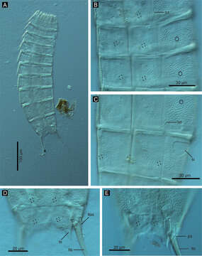
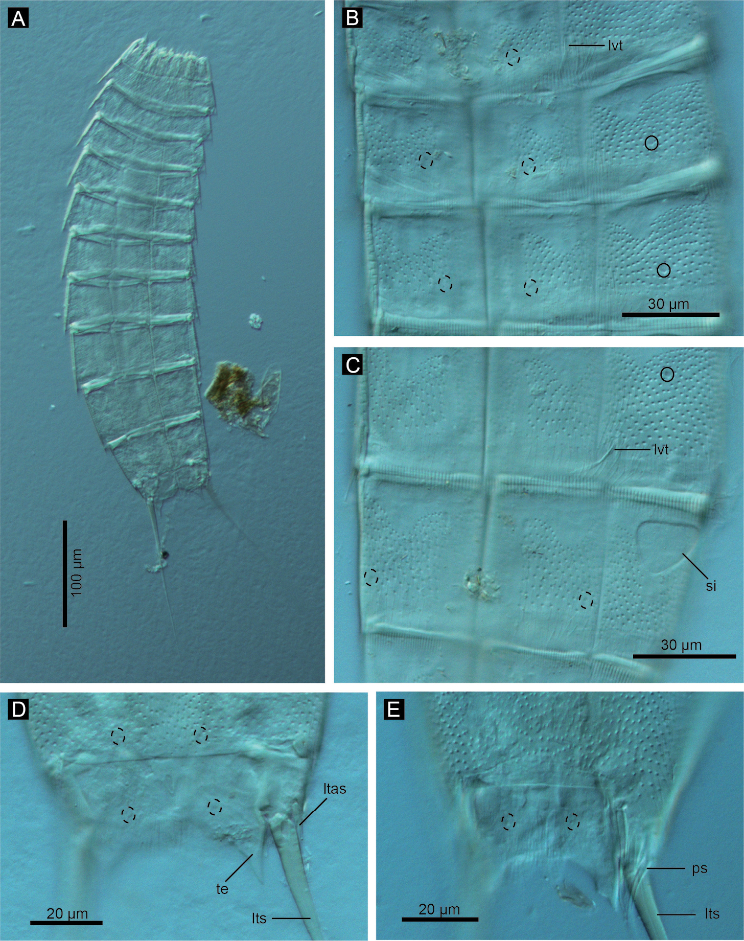
Figure 3.Echinoderes komatsui sp. n., Nomarski photomicrographs. A Entire animal B segments 5–7, ventral view C segments 8 and 9, ventral view D segments 10 and 11 of female, dorsal view E segments 10 and 11 of male, dorsal view. Complete circles indicate type 2 glandular cell outlet; dashed circles indicate sensory spots. Abbreviations: ltas, lateral terminal accessory spine; lts, lateral terminal spine; lvt, lateroventral tubule; ps, penile spine; si, sieve plate; te, tergal extension.
Included On The Following Pages:
- Eukaryota (eukaryotes)
- Opisthokonta (opisthokonts)
- Metazoa (animals)
- Bilateria
- Protostomia (protostomes)
- Ecdysozoa (ecdysozoans)
- Scalidophora
- Life
- Cellular
- Kinorhyncha (mud dragons)
- Cyclorhagida
- Echinoderidae
- Echinoderes
- Echinoderes komatsui
This image is not featured in any collections.
Source Information
- license
- cc-by-3.0
- copyright
- Hiroshi Yamasaki, Shinta Fujimoto
- bibliographic citation
- Yamasaki H, Fujimoto S (2014) Two new species in the Echinoderes coulli group (Echinoderidae, Cyclorhagida, Kinorhyncha) from the Ryukyu Islands, Japan ZooKeys 382: 27–52
- original
- original media file
- visit source
- partner site
- Zookeys
- ID