Image of Trigonalyidae

Description:
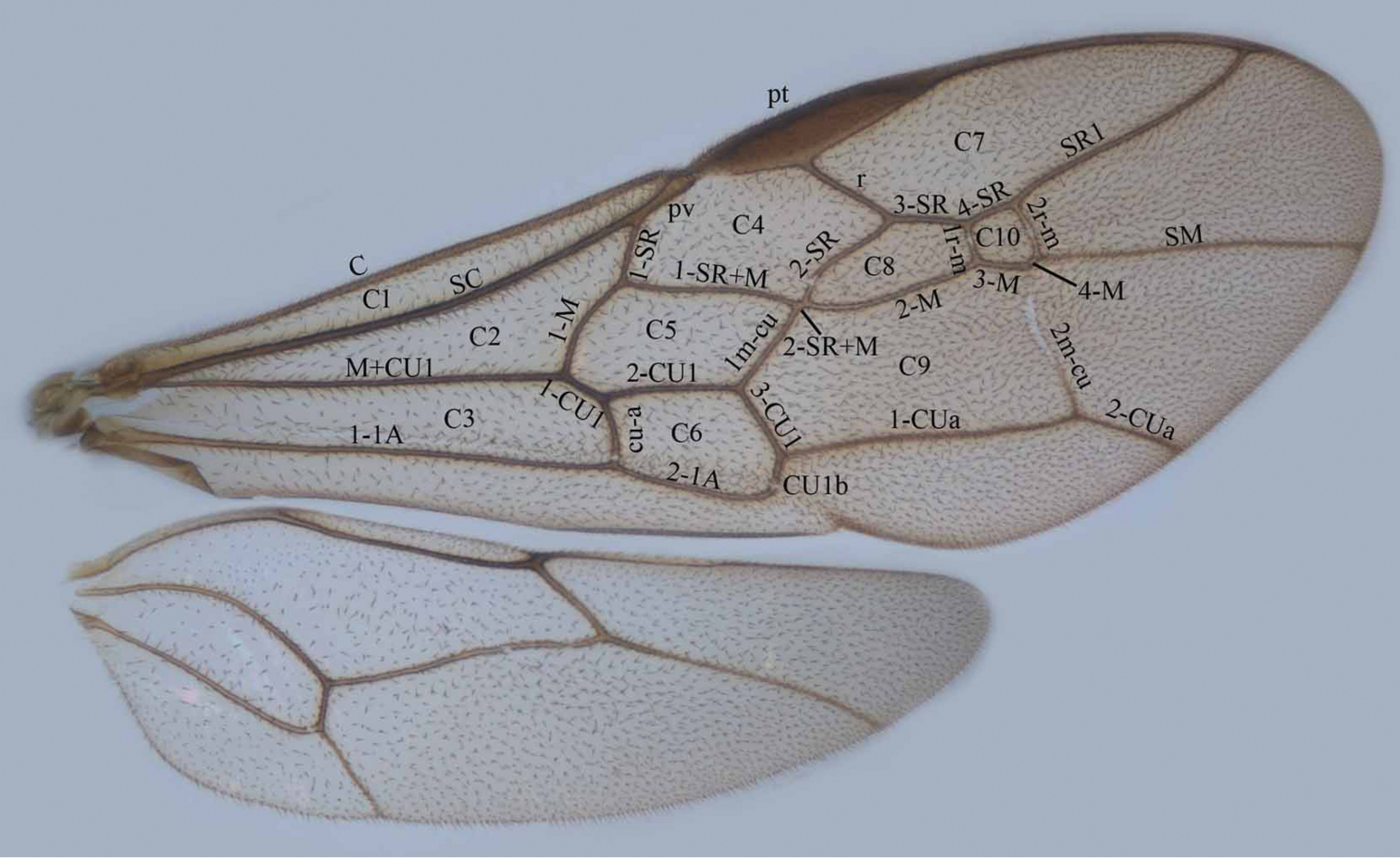
Figure 1.Wings of Trigonalyidae. C1 costal cell C2 medial cell C3 submedial cell C4 first submarginal cell C5 discal cell C6 subdiscal cell C7 marginal cell C8 second submarginal cell C9 second discal cell C10 third submarginal cell pt pterostigma pv parastigmal vein.
Included On The Following Pages:
This image is not featured in any collections.
Source Information
- license
- cc-by-3.0
- copyright
- Hua-yan Chen, Cornelis van Achterberg, Jun-hua He, Zai-fu Xu
- bibliographic citation
- Chen H, van Achterberg C, He J, Xu Z (2014) A revision of the Chinese Trigonalyidae (Hymenoptera, Trigonalyoidea) ZooKeys 385: 1–207
- original
- original media file
- visit source
- partner site
- Zookeys
- ID