Image of Eupoa lehtineni Logunov & Marusik 2014
Description:
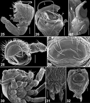
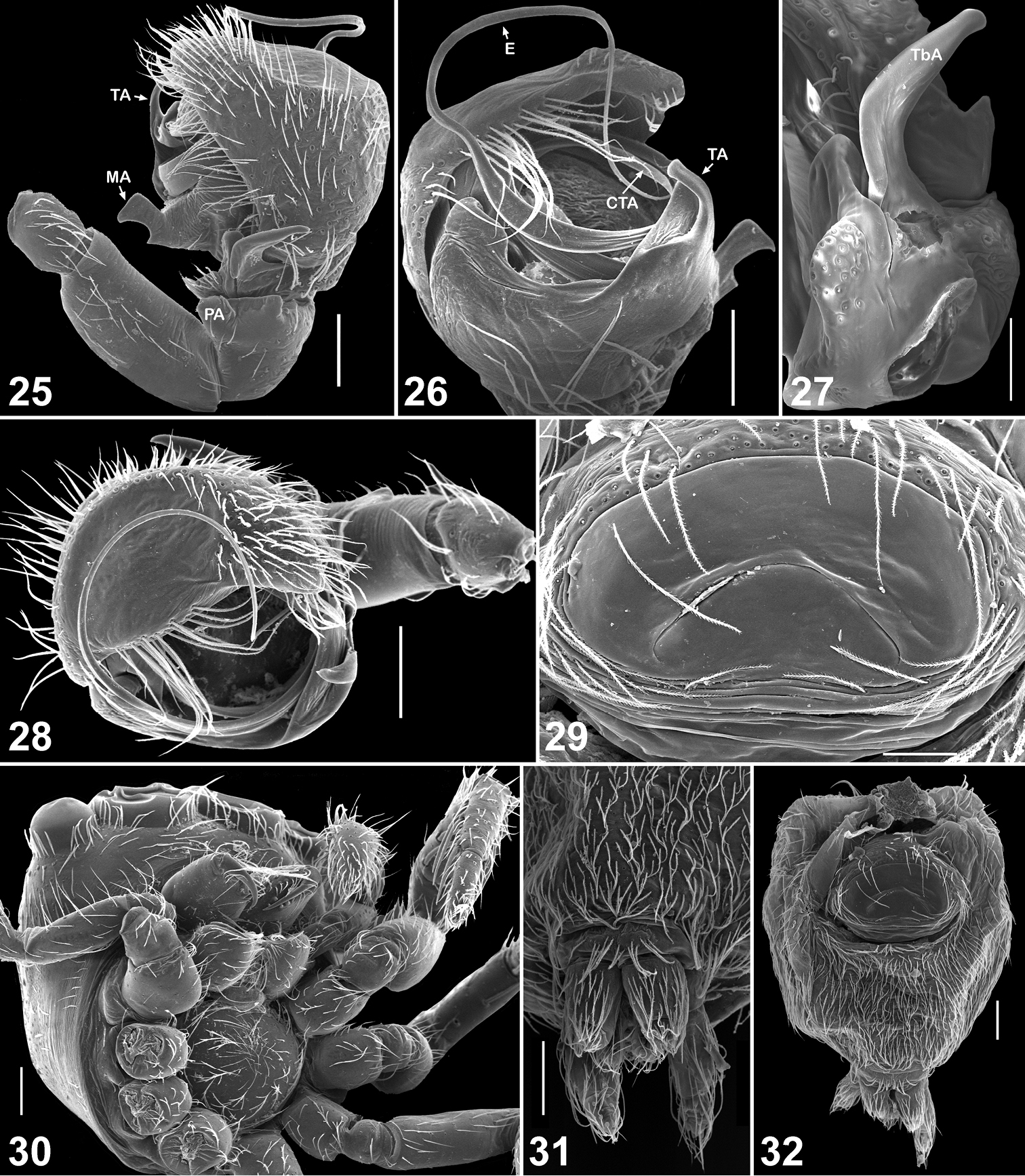
Figures 25–32.Copulatory organs and somatic characters of Eupoa lehtineni sp. n. 25 male palp, retrolateral view 26 ditto, median view 27 male tibial apophysis, retrolateral view 28 male palp, apical view 29 epigyne, ventral view 30 female carapace, ventral view 31 female spinnerets, ventral view 32 female abdomen, ventral view. Abbreviations as explained in ‘Material and methods’. Scale bars: 50 μm (27, 29), 0.1 mm (25–26, 28, 30–32).
Included On The Following Pages:
- Life
- Cellular
- Eukaryota (eukaryotes)
- Opisthokonta (opisthokonts)
- Metazoa (animals)
- Bilateria
- Protostomia (protostomes)
- Ecdysozoa (ecdysozoans)
- Arthropoda (arthropods)
- Chelicerata (chelicerates)
- Arachnida (arachnids)
- Araneae (spiders)
- Opisthothelae
- Araneomorphae
- Entelegynae
- Retrolateral tibial apophysis
- Salticidae (jumping spiders)
- Eupoa
- Eupoa lehtineni
- Panarthropoda
This image is not featured in any collections.
Source Information
- license
- cc-by-3.0
- copyright
- Dmitri V. Logunov, Yuri M. Marusik
- bibliographic citation
- Logunov D, Marusik Y (2014) Taxonomic notes on the genus Eupoa Żabka, 1985 (Arachnida, Araneae, Salticidae) ZooKeys 410: 63–93
- original
- original media file
- visit source
- partner site
- Zookeys
- ID