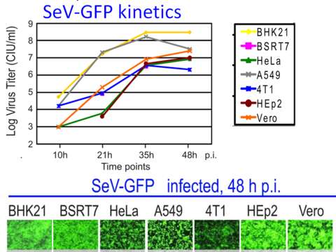
Sensitivity of different cell lines to Sendai virus infection
Description:
Description: English: The top panel shows one-step kinetics of viral replication in seven cell lines. Cells were infected with SeV-GFP at MOI of 3 CIU/cell (1 h absorption), washed 3 times with PBS, and kept in SFM. The media containing newly generated virions was collected at the indicated time points and viral titrations were performed on Vero cells (for SeV). The bottom panel shows photographs of seven cell lines infected with SeV-GFP at MOI 3 CIU/cell 48 hours post infection. Fluorescence microscopy images were captured at 10× magnification. Date: 22 June 2010. Source: Cell type mediated resistance of vesicular stomatitis virus and Sendai virus to ribavirin. PLoS One. 2010 Jun 22;5(6):e11265. doi: 10.1371/journal.pone.0011265. Author: Shah NR, Sunderland A, Grdzelishvili VZ.
Included On The Following Pages:
- Biota
- Virus
- Riboviria
- Orthornavirae
- Negarnaviricota
- Haploviricotina
- Monjiviricetes
- Mononegavirales
- Paramyxoviridae
- Paramyxovirinae
- Respirovirus
- Sendai virus
This image is not featured in any collections.
Source Information
- license
- cc-by-sa-3.0
- copyright
- Shah NR, Sunderland A, Grdzelishvili VZ.
- creator
- Shah NR, Sunderland A, Grdzelishvili VZ.
- source
- Cell type mediated resistance of vesicular stomatitis virus and Sendai virus to ribavirin.
- original
- original media file
- visit source
- partner site
- Wikimedia Commons
- ID