Viruses-12-00161-g006
Description:
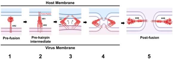
Summary.mw-parser-output table.commons-file-information-table,.mw-parser-output.fileinfotpl-type-information{border:1px solid #a2a9b1;background-color:#f8f9fa;padding:5px;font-size:95%;border-spacing:2px;box-sizing:border-box;margin:0;width:100%}.mw-parser-output table.commons-file-information-table>tbody>tr,.mw-parser-output.fileinfotpl-type-information>tbody>tr{vertical-align:top}.mw-parser-output table.commons-file-information-table>tbody>tr>td,.mw-parser-output table.commons-file-information-table>tbody>tr>th,.mw-parser-output.fileinfotpl-type-information>tbody>tr>td,.mw-parser-output.fileinfotpl-type-information>tbody>tr>th{padding:4px}.mw-parser-output.fileinfo-paramfield{background:#ccf;text-align:right;padding-right:0.4em;width:15%;font-weight:bold}.mw-parser-output.commons-file-information-table+table.commons-file-information-table,.mw-parser-output.commons-file-information-table+div.commons-file-information-table>table{border-top:0;padding-top:0;margin-top:-8px}@media only screen and (max-width:719px){.mw-parser-output table.commons-file-information-table,.mw-parser-output.commons-file-information-table.fileinfotpl-type-information{border-spacing:0;padding:0;word-break:break-word;width:100%!important}.mw-parser-output.commons-file-information-table>tbody,.mw-parser-output.fileinfotpl-type-information>tbody{display:block}.mw-parser-output.commons-file-information-table>tbody>tr>td,.mw-parser-output.commons-file-information-table>tbody>tr>th,.mw-parser-output.fileinfotpl-type-information>tbody>tr>td,.mw-parser-output.fileinfotpl-type-information>tbody>tr>th{padding:0.2em 0.4em;text-align:left;text-align:start}.mw-parser-output.commons-file-information-table>tbody>tr,.mw-parser-output.fileinfotpl-type-information>tbody>tr{display:flex;flex-direction:column}.mw-parser-output.commons-file-information-table+table.commons-file-information-table,.mw-parser-output.commons-file-information-table+div.commons-file-information-table>table{margin-top:-1px}.mw-parser-output.fileinfo-paramfield{box-sizing:border-box;flex:1 0 100%;width:100%}} Description: English: The SeV fusion protein (F) is a trimeric molecule belonging to class I viral membrane fusion protein. Each monomer of the trimer is synthesized as an F0 precursor. It must be cleaved by host protease into F1 and F2 subunits that remained connected through a disulfide linkage. Without such cleavage, the SeV virions are not infective. The cleavage site is located N-terminal to the fusion peptide which has N-(HR1) and C-(HR2) terminal heptad repeat domains. Only cleaved fusion protein (F) promotes the fusion of the virus envelope and host membranes. The pre-fusion, F protein is shown in red (1). Receptor-binding signals from the F-protein during the attachment process trigger the release of the fusion peptide, which inserts itself into the host cell membrane. The insertion of the fusion peptide parallels the conversion of the HR1 domain from a set of helical structures to a highly unstable, extended helical trimeric coil-coil domain (2). HR1 spirals attach F to the host cell membrane (3). Two lipid bilayers fuse with each other (4). The complete fusion of the HR2 and HR1 domains indicates the establishment of a six-helix bundle structure (6HB) (post-fusion F-structure shown in red), which leads to the pore formation and the completion of the fusion process (5). Date: 30 January 2020. Source: https://www.ncbi.nlm.nih.gov/pmc/articles/PMC7077268/. Author: Kristopher D. Azarm and Benhur Lee*.
Included On The Following Pages:
- Biota
- Virus
- Riboviria
- Orthornavirae
- Negarnaviricota
- Haploviricotina
- Monjiviricetes
- Mononegavirales
- Paramyxoviridae
- Paramyxovirinae
- Respirovirus
- Sendai virus
This image is not featured in any collections.
Source Information
- license
- cc-by-sa-3.0
- copyright
- Kristopher D. Azarm and Benhur Lee*
- creator
- Kristopher D. Azarm and Benhur Lee*
- source
- https://www.ncbi.nlm.nih.gov/pmc/articles/PMC7077268/
- original
- original media file
- visit source
- partner site
- Wikimedia Commons
- ID