Fmicb-07-01740-g004
Description:
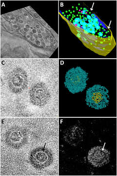
Description: English: Snapshots of electron tomograms and models of free and intracellular bacteriophages infecting Methylomirabilis cells. Tomogram (A) and model (B) of an infected Methylomirabilis cell. Most bacteriophages have the capsid (blue) assembled around the electron dense core (green). Some bacteriophages are still in the process of assembly and only consist of the electron dense core (pink). The cell is swollen and the cytoplasmic membrane (dark blue) is broken at many places (arrows). The cell wall (yellow) is still intact. All green electron dense cores were surrounded by a capsid, but not all capsids were modeled for reasons of clarity. Tomogram (C) and isosurface density model (D) of two free bacteriophages showing the icosahedral capsid (blue) and electron dense core (yellow). Tomogram (E) and Chimera model (F) showing two free bacteriophages. The electron dense core is enclosed by a putative membrane (arrows). Date: 8 November 2016. Source: Fig. 4 at https://www.frontiersin.org/articles/10.3389/fmicb.2016.01740/full Ultrastructure and Viral Metagenome of Bacteriophages from an Anaerobic Methane Oxidizing Methylomirabilis Bioreactor Enrichment Culture. In: Frontiers in Microbiology, volume 7 (2016), p1740, doi:10.3389/fmicb.2016.01740, ISSN 1664-302X . Author: Lavinia Gambelli, Geert Cremers, Rob Mesman, Simon Guerrero, Bas E. Dutilh, Mike S. M. Jetten, Huub J. M. Op den Camp, Laura van Niftrik. Other versions: .
Included On The Following Pages:
- Life (creatures)
- Cellular (cellular organisms)
- Bacteria
- Proteobacteria (Purple Bacteria & relatives)
This image is not featured in any collections.
Source Information
- license
- cc-by-sa-3.0
- copyright
- Lavinia Gambelli, Geert Cremers, Rob Mesman, Simon Guerrero, Bas E. Dutilh, Mike S. M. Jetten, Huub J. M. Op den Camp, Laura van Niftrik
- creator
- Lavinia Gambelli, Geert Cremers, Rob Mesman, Simon Guerrero, Bas E. Dutilh, Mike S. M. Jetten, Huub J. M. Op den Camp, Laura van Niftrik
- source
- Fig. 4 at https://www.frontiersin.org/articles/10.3389/fmicb.2016.01740/full Ultrastructure and Viral Metagenome of Bacteriophages from an Anaerobic Methane Oxidizing Methylomirabilis Bioreactor Enrichment Culture. In: Frontiers in Microbiology, volume 7 (2016), p1740, doi:10.3389/fmicb.2016.01740, ISSN 1664-302X
- original
- original media file
- visit source
- partner site
- Wikimedia Commons
- ID