Micrograph of Entamoeba histolytica
Description:
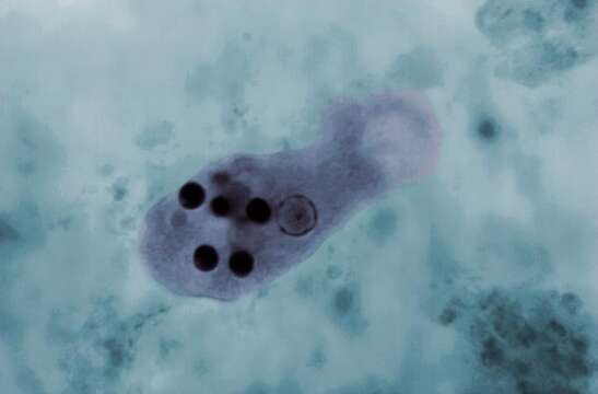
Summary.mw-parser-output table.commons-file-information-table,.mw-parser-output.fileinfotpl-type-information{border:1px solid #a2a9b1;background-color:#f8f9fa;padding:5px;font-size:95%;border-spacing:2px;box-sizing:border-box;margin:0;width:100%}.mw-parser-output table.commons-file-information-table>tbody>tr,.mw-parser-output.fileinfotpl-type-information>tbody>tr{vertical-align:top}.mw-parser-output table.commons-file-information-table>tbody>tr>td,.mw-parser-output table.commons-file-information-table>tbody>tr>th,.mw-parser-output.fileinfotpl-type-information>tbody>tr>td,.mw-parser-output.fileinfotpl-type-information>tbody>tr>th{padding:4px}.mw-parser-output.fileinfo-paramfield{background:#ccf;text-align:right;padding-right:0.4em;width:15%;font-weight:bold}.mw-parser-output.commons-file-information-table+table.commons-file-information-table,.mw-parser-output.commons-file-information-table+div.commons-file-information-table>table{border-top:0;padding-top:0;margin-top:-8px}@media only screen and (max-width:719px){.mw-parser-output table.commons-file-information-table,.mw-parser-output.commons-file-information-table.fileinfotpl-type-information{border-spacing:0;padding:0;word-break:break-word;width:100%!important}.mw-parser-output.commons-file-information-table>tbody,.mw-parser-output.fileinfotpl-type-information>tbody{display:block}.mw-parser-output.commons-file-information-table>tbody>tr>td,.mw-parser-output.commons-file-information-table>tbody>tr>th,.mw-parser-output.fileinfotpl-type-information>tbody>tr>td,.mw-parser-output.fileinfotpl-type-information>tbody>tr>th{padding:0.2em 0.4em;text-align:left;text-align:start}.mw-parser-output.commons-file-information-table>tbody>tr,.mw-parser-output.fileinfotpl-type-information>tbody>tr{display:flex;flex-direction:column}.mw-parser-output.commons-file-information-table+table.commons-file-information-table,.mw-parser-output.commons-file-information-table+div.commons-file-information-table>table{margin-top:-1px}.mw-parser-output.fileinfo-paramfield{box-sizing:border-box;flex:1 0 100%;width:100%}} Description: English: This photomicrograph of a trichrome-stained specimen revealed the presence of an Entamoeba histolytica trophozoite, within which a number of phagocytized erythrocytes could be seen as dark, round inclusions. Date: 1 January 1966. Source: CDC/ Dr. Mae Melvin; Dr. Greene (1966). Entamoeba histolytica. Public Health Image Library (PHIL) at Centers for Disease Control and Prevention (CDC) at the U.S. Department of Health & Human Services. - "Copyright Restrictions:None - This image is in the public domain and thus free of any copyright restrictions.". Author: CDC/ Dr. Mae Melvin; Dr. Greene. Other versions: Annotated.
Included On The Following Pages:
- Life (creatures)
- Cellular (cellular organisms)
- Eukaryota (eukaryotes)
- Amoebozoa (amoeboid protists)
- Evosea
- Archamoebae
- Mastigamoebida
- Entamoebidae
- Entamoeba
- Entamoeba histolytica
This image is not featured in any collections.
Source Information
- license
- cc-publicdomain
- creator
- CDC/ Dr. Mae Melvin; Dr. Greene|year=1966 - "Copyright Restrictions:None - This image is in the public domain and thus free of any copyright restrictions."
- source
- CDC/ Dr. Mae Melvin; Dr. Greene (1966). Entamoeba histolytica. Public Health Image Library (PHIL) at Centers for Disease Control and Prevention (CDC) at the U.S. Department of Health & Human Services. - "Copyright Restrictions:None - This image is in the public domain and thus free of any copyright restrictions."
- original
- original media file
- visit source
- partner site
- Wikimedia Commons
- ID