Trichomonas vaginalis phase contrast microscopy
Description:
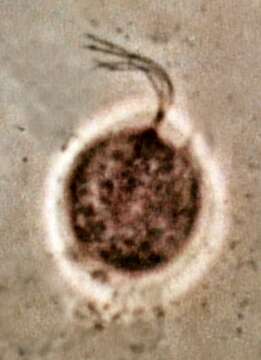
Description: English: Trichomonas vaginalis photographed by phase contrast microscopy. Date: 17 July 2010. Source: Own work. Author: Dr Graham Beards.
Included On The Following Pages:
- Life
- Cellular
- Eukaryota (eukaryotes)
- Excavates (excavates)
- Metamonada (metamonad)
- Parabasalia (parabasalids)
- Trichomonadida
- Trichomonadidae
- Trichomonas
- Trichomonas vaginalis
This image is not featured in any collections.
Source Information
- license
- cc-by-sa-3.0
- copyright
- Dr Graham Beards
- creator
- Dr Graham Beards
- original
- original media file
- visit source
- partner site
- Wikimedia Commons
- ID