PhycRes-pre12392-fig-0001-m-Vampirovibrio-chlorellavorus
Description:
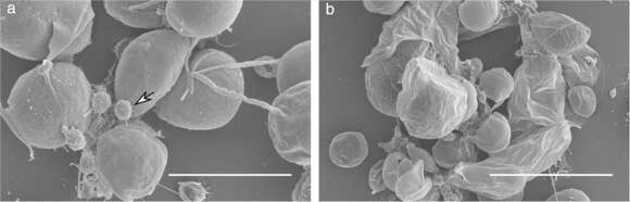
Description: English: Scanning Electron Micrographs of Chlorella sorokiniana and attached Vampirovibrio chlorellavorus cells. Images of samples collected from Arizona test site at (a) 10 000 × magnification with V. chlorellavorus indicated by the white arrow and (b) 5000 × magnification.Scale bars are displayed in white representing (a) 5.0 μm and (b) 10.0 μm. Date: 17 July 2019. Source: Fig. 1 at https://onlinelibrary.wiley.com/doi/10.1111/pre.12392 Vampirovibrio chlorellavorus draft genome sequence, annotation, and preliminary characterization of pathogenicity determinants Phycological Research Vol. 68, No. 1 p. 23-29, doi:10.1111/pre.12392 . Author: Blake T. Hovde, Seth A. Steichen, Shawn R. Starkenburg, Judith K. Brown. Other versions: This file has an extracted image: File:PhycRes-pre12392-fig-0001a-m-Vampirovibrio-chlorellavorus.jpg..
Included On The Following Pages:
- Life
- Cellular
- Eukaryota (eukaryotes)
- Archaeplastida (plants)
- Chloroplastida
- Chlorophyta (chlorophytes)
- Trebouxiophyceae
- Chlorellales
- Chlorellaceae
- Chlorella
This image is not featured in any collections.
Source Information
- license
- cc-by-sa-3.0
- copyright
- Blake T. Hovde, Seth A. Steichen, Shawn R. Starkenburg, Judith K. Brown
- creator
- Blake T. Hovde, Seth A. Steichen, Shawn R. Starkenburg, Judith K. Brown
- source
- Fig. 1 at https://onlinelibrary.wiley.com/doi/10.1111/pre.12392 Vampirovibrio chlorellavorus draft genome sequence, annotation, and preliminary characterization of pathogenicity determinants Phycological Research Vol. 68, No. 1 p. 23-29, doi:10.1111/pre.12392
- original
- original media file
- visit source
- partner site
- Wikimedia Commons
- ID