Schistosoma 20041-300
Description:
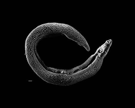
Description: English: Electron micrograph of an adult male Schistosoma parasite worm. The bar (bottom left) represents a magnification of 500 μm. Français : Photographie au micrographe électronique d'un ver parasite Schistosoma adulte mâle. La barre (en bas à gauche) représente une longueur de 500 μm. Date: 2 September 2009. Source: http://www.genome.gov/dmd/img.cfm?node=Photos/Animals/Trihinella (NHGRI-79094.jpg, formerly 20041-300.jpg) Transferred from en.wikipedia to Commons by User:Magnus Manske using CommonsHelper. Author: David Williams, Illinois State University. Permission(Reusing this file): Page states "This image is freely available and may be used without special permission." See also http://www.genome.gov/copyright.cfm.
Included On The Following Pages:
- Life (creatures)
- Cellular (cellular organisms)
- Eukaryota (eukaryotes)
- Opisthokonta (opisthokonts)
- Metazoa (Animal)
- Bilateria
- Protostomia (protostomes)
- Ecdysozoa (ecdysozoans)
- Arthropoda (arthropods)
- Pancrustacea
- Multicrustacea (typical crustaceans)
- Malacostraca (malacostracans)
- Eumalacostraca
- Peracarida (peracarids)
- Isopoda (isopods)
- Asellota
- Janiroidea
- Mictosomatidae
- Mictosoma
This image is not featured in any collections.
Source Information
- license
- cc-publicdomain
- creator
- David Williams, Illinois State University
- source
- http://www.genome.gov/dmd/img.cfm?node=Photos/Animals/Trihinella (NHGRI-79094.jpg, formerly 20041-300.jpg)
- original
- original media file
- visit source
- partner site
- Wikimedia Commons
- ID