Yersinia pestis HHS
Description:
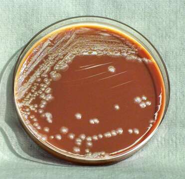
Description: English: This photograph depicts the colonial morphology displayed by Gram-negative Yersinia pestis bacteria, which was grown on a medium of chocolate agar, for a 72 hour time period, at a temperature of 25°C. Note that PHIL 12470 displays a closer view of these Y. pestis bacterial colonies. See also PHIL 12468 for the appearance of Y. pestis colonies grown on the same medium, and at the same time temperature, but for a lesser time period (48hr); PHIL 12471 displaying growth on the same medium, but for a lesser time period (48hr), and at a greater temperature (37°C); PHIL 12472 displaying growth on the same medium, for the same amount of time, but at a greater temperature (37°C); PHIL 12473 displaying Y. pestis colonies grown on the same medium and temperature, but at one-third the time (24hr). Date: 2010. Source: : This media comes from the Centers for Disease Control and Prevention's Public Health Image Library (PHIL), with identification number #12469. Note: Not all PHIL images are public domain; be sure to check copyright status and credit authors and content providers. العربية | Deutsch | English | македонски | slovenščina | +/−. Author: Department of Health and Human Services.
Included On The Following Pages:
- Life
- Cellular
- Bacteria
- Proteobacteria (Purple Bacteria & relatives)
- Gammaproteobacteria
- Enterobacterales
- Yersiniaceae
- Yersinia
- 'Yersinia pseudotuberculosis complex'
- Yersinia pestis
This image is not featured in any collections.
Source Information
- license
- cc-publicdomain
- creator
- Department of Health and Human Services
- source
- {{CDC-PHIL|12469}}
- original
- original media file
- visit source
- partner site
- Wikimedia Commons
- ID