1999 Escherichia-coli
Description:
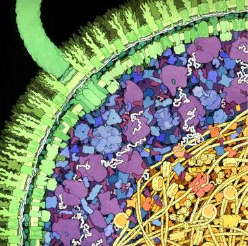
Description: English: This illustration shows a cross-section of a small portion of an Escherichia coli cell. The cell wall, with two concentric membranes studded with transmembrane proteins, is shown in green. A large flagellar motor crosses the entire wall, turning the flagellum that extends upwards from the surface. The cytoplasmic area is colored blue and purple. The large purple molecules are ribosomes and the small, L-shaped maroon molecules are tRNA, and the white strands are mRNA. Enzymes are shown in blue. The nucleoid region is shown in yellow and orange, with the long DNA circle shown in yellow, wrapped around HU protein (bacterial nucleosomes). In the center of the nucleoid region shown here, you might find a replication fork, with DNA polymerase (in red-orange) replicating new DNA. Date: 1999. Source: https://pdb101.rcsb.org/sci-art/goodsell-gallery/escherichia-coli. Author: David Goodsell.
Included On The Following Pages:
- Life (creatures)
- Cellular (cellular organisms)
- Bacteria
- Proteobacteria (Purple Bacteria & relatives)
- Gammaproteobacteria
- Enterobacterales
- Enterobacteriaceae
- Escherichia
- Escherichia coli (E. coli)
This image is not featured in any collections.
Source Information
- license
- cc-by-3.0
- copyright
- David Goodsell
- creator
- David Goodsell
- source
- https://pdb101.rcsb.org/sci-art/goodsell-gallery/escherichia-coli
- original
- original media file
- visit source
- partner site
- Wikimedia Commons
- ID