Doi.10.1128.JVI.00175-20.F2.large
Description:
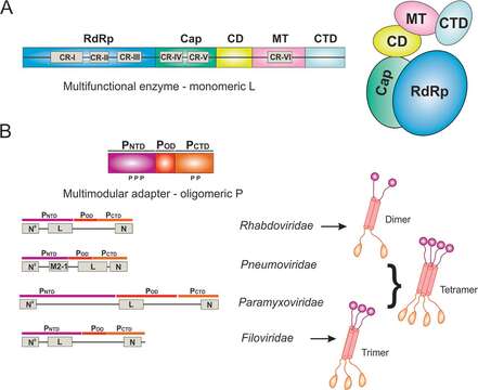
Description: English: The domain organization and architecture of L and P. (A) The domain organization and cartoon representation of the multifunctional enzyme monomeric L. The conserved regions (CRs) I to VI are labeled in gray boxes. The RNA-dependent RNA polymerization domain (RdRp), capping domain (Cap), connector domain (CD), methyltransferase domain (MT), and C-terminal domain (CTD) of L are colored in blue, green, yellow, pink, and cyan, respectively. (B) The domain organization and cartoon representation of the multimodular adapter oligomeric P. The intrinsically disordered N-terminal domain (PNTD), oligomerization domain (POD), and C-terminal domain (PCTD) are colored in magenta, red, and orange, respectively. The interaction regions with other viral proteins, including L, N, RNA-free N (N0), and accessory protein (M2-1), are labeled in gray boxes. The representative P oligomers are shown for the representative virus families Rhabdoviridae, Pneumoviridae, Paramyxoviridae, and Filoviridae. Date: 27 October 2020. Source: Liang B. 2020. Structures of the Mononegavirales polymerases. J Virol 94:e00175-20. https://doi.org/10.1128/JVI.00175-20. Author: Bo Liang.
Included On The Following Pages:
- Biota
- Virus
- Riboviria
- Orthornavirae
- Negarnaviricota
- Haploviricotina
- Monjiviricetes
- Mononegavirales
- Rhabdoviridae (rabies virus and relatives)
- Lyssavirus
- Rabies virus
This image is not featured in any collections.
Source Information
- license
- cc-by-3.0
- copyright
- Bo Liang
- original
- original media file
- visit source
- partner site
- Wikimedia Commons
- ID