Doi.10.1128.JVI.00175-20.F3.large
Description:
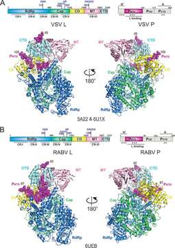
Description: English: The cryo-EM structures of the Rhabdoviridae polymerases. (A) Linear domain representation of the L and P proteins of the vesicular stomatitis virus (VSV) polymerase. The cartoon view of 3.8-Å (PDB: 5A22) and 3.0-Å (PDB: 6U1X) cryo-EM structures of the VSV polymerase are shown. (B) Linear domain representation of the L and P proteins of the rabies virus (RABV) polymerase. The cartoon view of the 3.3-Å (PDB: 6UEB) cryo-EM structure of the RABV polymerase is shown. The RNA-dependent RNA polymerization domain (RdRp), capping domain (Cap), connector domain (CD), methyltransferase domain (MT), C-terminal domain (CTD) of L, and PNTD are colored in blue, green, yellow, pink, cyan, and magenta, respectively. The missing domains are colored in gray. The PNTD is highlighted as spheres, and the terminal residue numbers of the modeled P segments are indicated. The PDB accession codes are underlined. Date: 27 October 2020. Source: Liang B. 2020. Structures of the Mononegavirales polymerases. J Virol 94:e00175-20. https://doi.org/10.1128/JVI.00175-20. Author: Bo Liang.
Included On The Following Pages:
- Biota
- Virus
- Riboviria
- Orthornavirae
- Negarnaviricota
- Haploviricotina
- Monjiviricetes
- Mononegavirales
- Rhabdoviridae (rabies virus and relatives)
- Lyssavirus
- Rabies virus
This image is not featured in any collections.
Source Information
- license
- cc-by-3.0
- copyright
- Bo Liang
- original
- original media file
- visit source
- partner site
- Wikimedia Commons
- ID