Doi.10.1128.JVI.00175-20.F7.large
Description:
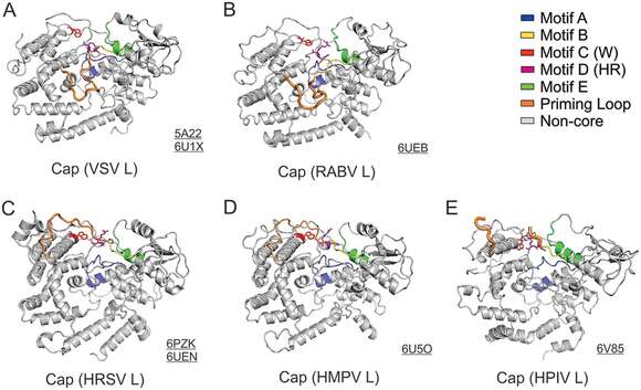
Description: English: Structural comparison of the Cap domain. The motifs A to E of the Cap domain of the Rhabdoviridae (VSV and RABV), Pneumoviridae (HRSV and HMPV), and Paramyxoviridae (HPIV) L are shown as ribbon diagrams in blue, yellow, red, magenta, and green, respectively. Those motifs are centered around the active site motif D (HR). The proposed priming loop (orange) is next to motif B. The PDB accession codes are underlined. Date: 27 October 2020. Source: Liang B. 2020. Structures of the Mononegavirales polymerases. J Virol 94:e00175-20. https://doi.org/10.1128/JVI.00175-20. Author: Bo Liang.
Included On The Following Pages:
- Biota
- Virus
- Riboviria
- Orthornavirae
- Negarnaviricota
- Haploviricotina
- Monjiviricetes
- Mononegavirales
- Rhabdoviridae (rabies virus and relatives)
- Lyssavirus
- Rabies virus
This image is not featured in any collections.
Source Information
- license
- cc-by-3.0
- copyright
- Bo Liang
- original
- original media file
- visit source
- partner site
- Wikimedia Commons
- ID