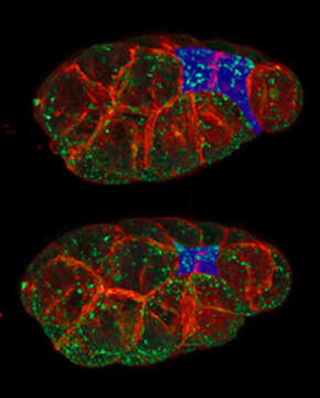
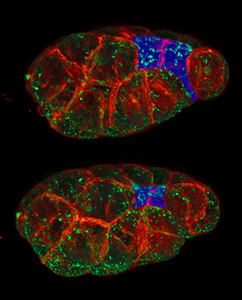
Apical Constriction
Description:
Description: English: Two stages in the constriction of the apical surfaces (blue) of a pair of cells in a C. elegans embryo. Plasma membranes (red) and myosin (green) are marked. Goldstein lab, UNC Chapel Hill. Date: 23 January 2012, 09:00:36. Source: Own work. Author: Rpgch.
Included On The Following Pages:
- Life
- Cellular
- Eukaryota (eukaryotes)
- Opisthokonta (opisthokonts)
- Metazoa (animals)
- Bilateria
- Protostomia (protostomes)
- Ecdysozoa (ecdysozoans)
- Nematoda (nematodes)
- Chromadorea
- Chromadoria
- Rhabditida (rhabditid)
- Rhabditina
- Rhabditomorpha
- Rhabditoidea
- Rhabditidae
- Caenorhabditis
- Caenorhabditis elegans
This image is not featured in any collections.
Source Information
- license
- cc-by-sa-3.0
- copyright
- Rpgch
- creator
- Rpgch
- original
- original media file
- visit source
- partner site
- Wikimedia Commons
- ID