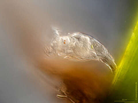
Image of Philodina megalotrocha Ehrenberg 1832
Description:
Philodina megalotrocha We see the foot-tail with the two spurs. The foot is partially retracted, which becomes visible through the annulated integument. Eyespots and trophi are also visible. Scale bar indicates 50 µm. Please press the MORE button for skipping to the annotated version.Sample from a tropical freshwater aquarium. Sampling date 3/2023. The image was built up using several photomicrographic frames with manual stacking technique. Images were taken using Zeiss Axioplan with Olympus OM-D M5 MKII. Image under Creative Commons License V 3.0 (CC BY-NC-SA). Place name: Tropical freshwater aquarium Latitude: 54.3018013 Longitude: 10.07120132 Das Bild zeigt den Fuß mit den Sporen. Der Fuß ist teilweise eingezogen, was das geringelte Integument anzeigt. Augenflecken und Kauer sind ebenfalls sichtbar. Der Messbalken markiert eine Länge von 50 µm. Bitte drücken Sie die Schaltfläche MORE, um zur kommentierten Version zu gelangen.Probe aus einem Süßwasseraquarium. Datum der Aufsammlung: 3/2023. Mikrotechnik: Zeiss Axioplan, Kamera: Olympus OM-D M5 MKII. Creative Commons License V 3.0 (CC BY-NC-SA). For permission to use of (high-resolution) images please contact postmaster@protisten.de.
Included On The Following Pages:
- Life
- Cellular
- Eukaryota (eukaryotes)
- Opisthokonta (opisthokonts)
- Metazoa (animals)
- Bilateria
- Protostomia (protostomes)
- Spiralia (spiralians)
- Gnathifera
- Syndermata
- Rotifera (rotifers)
- Bdelloidea
- Philodinidae
- Philodina
- Philodina megalotrocha
This image is not featured in any collections.
Source Information
- license
- cc-by-nc-sa-3.0
- copyright
- Wolfgang Bettighofer
- creator
- Wolfgang Bettighofer [email]
- original
- original media file
- visit source
- partner site
- protisten.de
- ID
_NEW.jpg)