Oral infraciliatue
Description:
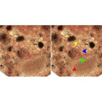
Anterior apical view of the infraciliature of Vasicola ciliata (Tatem, 1869). The cytostome is at the base of a shallow funnel-shaped depressin. There are three groups of cilia encircling the cytostome 1)the circumoral kinety (yellow arrowhead) is composed of a single row of transversely oriented dikinetids 2) the anterior row of "membranelles" composed of a single row of longitudinally oriented dikinetids (blue arrowhead 3) the posterior row of "membranelles", a band of four longitudinal orietd kinetids (green arrowhead). The second somatic kinety is indicated by the red arrowhead. Longitudinal fibrils extend from the cytostome to the right of the kinetids. Stained by the silver carbonate technique (see Foissner, W. Europ. J. Protistol., 27:313-330;1991). Brightfield.
Included On The Following Pages:
- Life
- Cellular
- Eukaryota (eukaryotes)
- SAR (Stramenopiles, Alveolates, Rhizaria)
- Alveolata (alveolates)
- Ciliophora (ciliates)
- Intramacronucleata
- Prostomatea
- Prostomatida
- Metacystidae
- Vasicola
- Vasicola ciliata
This image is not featured in any collections.
Source Information
- license
- cc-by-nc
- author
- William Bourland
- provider
- micro*scope
- original
- original media file
- visit source
- partner site
- micro*scope
- ID