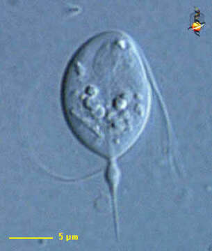
portrait, DIC
Description:
Foaina (foe-een-a) is one of the trichomonad flagellates - mostly endobiotic with four flagella arising from a point near the front of the cell. Three anterior free flagella and a recurrent one adhering to the cell body only on its proximal part. Three flagella are tightly grouped together, separating only at their distal ends. Usually with several cytoskeletal structures, an axial axostyle made of microtubules which encloses the nucleus anteriorly, as well as a costa which often lies under one of the flagella. The axostyle projects from the posterior end of the cell and may be adapted to form a holdfast which attaches the cell to a piece of debris. Parabasal body (dictyosomes) rod-, disc- or V-shaped. Electron microscopy has shown the trichomonad characters and particularly the presence of an infrakinetosomal body similar to that of Tritrichomonas. About 20 species living in the intestinal tract of vertebrates. other species live in the gut of invertebrates, especially arthropods such as termites and roaches, coleoptera, tipulid larvae and myriapods. Nucleus evident near front of cell, axostyle extending along the axis of the cell and out of the posterior end. From the termite Cryptotermes. Differential interference contrast.
Included On The Following Pages:
- Life
- Cellular
- Eukaryota (eukaryotes)
- Excavates (excavates)
- Metamonada (metamonad)
- Parabasalia (parabasalids)
- Cristamonadida
- Foaina
This image is not featured in any collections.
Source Information
- license
- cc-by-nc
- author
- Linda Amaral Zettler, Lorraine Olendzenski and David Patterson
- provider
- micro*scope
- original
- original media file
- visit source
- partner site
- micro*scope
- ID