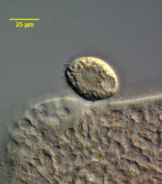
Sphaerophrya infesting Bursaria
Description:
Sphaerophrya insolita (Jankowski, 1973) infesting the large colpodid ciliate, Bursaria truncatella (Muller, 1773). The Sphaerophrya cells are ellipsoid and approximately 35 u in diameter. Sphaerophrya is thought to have lost its stalk during the transition to a parasitic mode of existence. The cells have capitate tentacles by which they adhere to the pellicle of the host cell (several of these are visible on the viewer's left). There is a central ellipsoid granular nucleus seen well here (the micronuclei have not been characterized). There is a single peripheral contractile vacuole (seen well in this cell). These individuals were found on B. truncatella collected from a temporary rainwater pool containing decaying grass near Boise, Idaho March 2005. DIC.
Included On The Following Pages:
- Life
- Cellular
- Eukaryota (eukaryotes)
- SAR (Stramenopiles, Alveolates, Rhizaria)
- Alveolata (alveolates)
- Ciliophora (ciliates)
- Intramacronucleata
- Phyllopharyngea
- Suctoria
- Exogenida
- Podophryidae
- Sphaerophrya
- Sphaerophrya insolita
This image is not featured in any collections.
Source Information
- license
- cc-by-nc
- author
- William Bourland
- provider
- micro*scope
- original
- original media file
- visit source
- partner site
- micro*scope
- ID