Surface detail
Description:
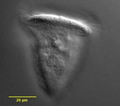
Surface detail of the peritrich ciliate, Pseudovorticella chlamydophora (Penard, 1922) Jankowski, 1976. Pseudovorticella is distinguished from Vorticella by silver staining which reveals a lattice-like silver line system in the former and circumferential lines without vertical connections in the latter. Pseudovorticella also has two contractile vacuoles. P. chlamydophora is distinguished by a distinct hyaline layer consisting of large cuboid pellicular blebs. The lattice-like pattern of these blebs is visible here. Feeds on bacteria. From freshwater pond near Boise, Idaho. DIC.
Included On The Following Pages:
- Life
- Cellular
- Eukaryota (eukaryotes)
- SAR (Stramenopiles, Alveolates, Rhizaria)
- Alveolata (alveolates)
- Ciliophora (ciliates)
- Intramacronucleata
- Oligohymenophorea
- Peritrichia
- Sessilida
- Vorticellidae
- Pseudovorticella
- Pseudovorticella chlamydophora
This image is not featured in any collections.
Source Information
- license
- cc-by-nc
- author
- William Bourland
- provider
- micro*scope
- original
- original media file
- visit source
- partner site
- micro*scope
- ID