Ventral view
Description:
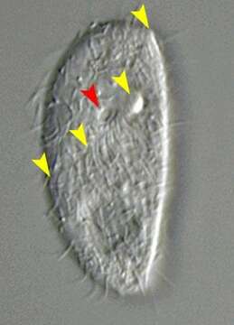
Portrait of the nassophorean ciliate, Chilodontopsis depressa (Perty, 1852). The cell is dorsoventrally flattened. The right side is convex meeting the straight left side at a rostrum. The somatic ciliature is denser on the ventral side with an indistinct feathery hypostomial frange of longer cilia slanting posteriorly to the cytostome from the rostrum anteriorly to the right margin of the body (yellow arrowheads). The right ventral kineties curve around the anterior end to meet the straight left ventral kineties to the left of the cytostome (red arrowhead) at the line of the hypostomial frange. The cytopharyngeal basket or cyrtos is seen anteriorly (red arrowhead). A distinctive large contractile vacuole fills the posterior end of the cell. The central macronucleus and micronucleus are spherical. C. depressa feeds on bacteria, diatoms and green algae. Collected from freshwater pond near Boise, Idaho September 2003. DIC optics
Included On The Following Pages:
- Life
- Cellular
- Eukaryota (eukaryotes)
- SAR (Stramenopiles, Alveolates, Rhizaria)
- Alveolata (alveolates)
- Ciliophora (ciliates)
- Intramacronucleata
- Nassophorea
- Synhymeniida
- Scaphidiodontidae
- Chilodontopsis
- Chilodontopsis depressa
This image is not featured in any collections.
Source Information
- license
- cc-by-nc
- author
- William Bourland
- provider
- micro*scope
- original
- original media file
- visit source
- partner site
- micro*scope
- ID