portrait
Description:
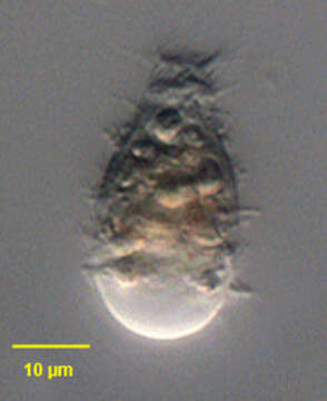
Portrait of Metacystis recurva (Penard,1922), a prostomatid ciliate. This individual has fled its lorica and is swimming free. In the lorica the body is usually elongate but quite contractile. The free-swimming individuals are typically contracted. The anterior is bluntly truncate and the posterior broader and rounded. The distinctive large, clear, protuberant posterior vacuole is seen in this image but this may sometimes be lacking leading to confusion with the similar genus, Vasicola. The oral aperture is apical, surrounded by four rows of peribuccal cilia. The kinetids of the longitudinal kineties line up with one another to form horizontal rows called paratenes. The cell surface may be transversely furrowed along these paratenes. There is often a long laterally located posterior cilium (not seen here). The central macronucleus and posterior contractile vacuole are not well seen here. The lorica is a narrow curved truncate cone shape with 12-15 transverse corrugations (thanks to Martin Kreutz for his translation of Kahlâs species description). Metacystis is said to feed on sulfur bacteria. From sapropelic freshwater aquaculture tank near Boise, Idaho. DIC optics.
Included On The Following Pages:
- Life
- Cellular
- Eukaryota (eukaryotes)
- SAR (Stramenopiles, Alveolates, Rhizaria)
- Alveolata (alveolates)
- Ciliophora (ciliates)
- Intramacronucleata
- Prostomatea
- Prostomatida
- Metacystidae
- Metacystis
- Metacystis recurva
This image is not featured in any collections.
Source Information
- license
- cc-by-nc
- author
- William Bourland
- provider
- micro*scope
- original
- original media file
- visit source
- partner site
- micro*scope
- ID