Anterior detail
Description:
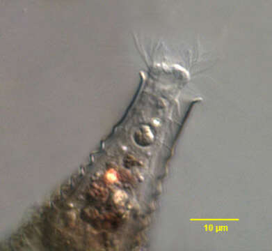
Anterior detail of Metacystis recurva (Penard,1922), a prostomatid ciliate. The oral aperture is apical, surrounded by four rows of long peribuccal cilia. The kinetids of the longitudinal kineties line up with one another to form horizontal rows called paratenes. The cell surface may be transversely furrowed along these paratenes. The highly refractile material in the neck of the cell is an aggregate of cytoplasmic crystals. The lorica is a narrow curved truncate cone shape with 12-15 transverse corrugations (thanks to Martin Kreutz for his translation of Kahlâs species description). The lorica is nearly colorless in young individuals and becomes sepia color with age, presumably due to deposition of minerals. The overlying cladocercan shell distorts the color in this image. Loricae are often found inside the vacant shells of cladocercans. ). Metacystis is said to feed on sulfur bacteria. From sapropelic freshwater aquaculture tank near Boise, Idaho. DIC optics.
Included On The Following Pages:
- Life
- Cellular
- Eukaryota (eukaryotes)
- SAR (Stramenopiles, Alveolates, Rhizaria)
- Alveolata (alveolates)
- Ciliophora (ciliates)
- Intramacronucleata
- Prostomatea
- Prostomatida
- Metacystidae
- Metacystis
- Metacystis recurva
This image is not featured in any collections.
Source Information
- license
- cc-by-nc
- author
- William Bourland
- provider
- micro*scope
- original
- original media file
- visit source
- partner site
- micro*scope
- ID