in vivo;right lateral view
Description:
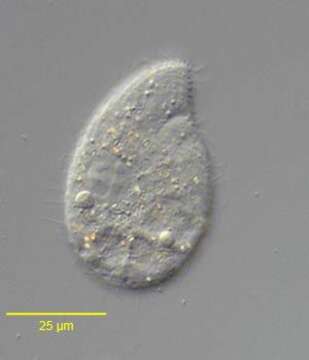
Right lateral view of the colpodid ciliate, Exocolpoda augustini (Foissner, 1987) Foissner, Agatha and Berger, 2002. Foissner erected the family Exocolpodidae based on the life cycle of its members, namely, cell division in free-swimming individuals instead of reproduction in division cysts as seen in the Colpodidae. He felt this life cycle characteristic,the unique boomerang-shaped left oral polykinetid and the unique thick-walled resting cyst of this species warranted its transfer to the new genus, Exocolpoda. The anterior of the cell is cone-shaped and the posterior globular.The small cytostome is in the anterior 1/4 of the cell.There are 25-35 somatic kineties composed of doubly ciliated dikinetids.The right somatic kineties spiral slightly on the long axis to end on the short preoral suture. The left kineties curve more strongly to perpendicularly abut the suture.There are two oral poykinetids. The lekt polykinetid has a unique angulated shape like a boomerang.The macronucleus is spherical.The nucleolus is ribbon-like.There is a single posterior contractile vacuole with a solitary excretory pore.Collected near Boise, Idaho (43°38'21.10"N 116°11'10.78"W elev. 2908 ft.) from an ice-covered temporary puddle containing leaf litter and dead grass.November, 2005.DIC.
Included On The Following Pages:
- Life
- Cellular
- Eukaryota (eukaryotes)
- SAR (Stramenopiles, Alveolates, Rhizaria)
- Alveolata (alveolates)
- Ciliophora (ciliates)
- Intramacronucleata
- Colpodea
- Colpodida
- Colpodidae
- Exocolpoda
- Exocolpoda augustini
This image is not featured in any collections.
Source Information
- license
- cc-by-nc
- author
- William Bourland
- provider
- micro*scope
- original
- original media file
- visit source
- partner site
- micro*scope
- ID