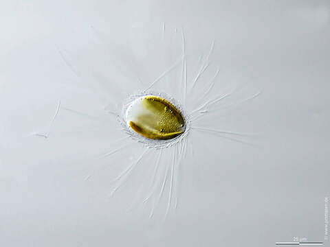
Image of Mallomonas caudata
Description:
Sampling date 11/2016. Scale bars indicate 25 µm (1–4), 10 µm (5).
Five images with different focal planes (four of them in a slide changer). Cells are covered by siliceous scales with appendages.
Please click on < or > on the image edges or on the dots at the bottom edge of the images to browse through the slides!
Place name: Lake Schierensee near Kiel (Schleswig-Holstein, Germany)
Latitude: 54.26256721 Longitude: 9.98369694
Microscope Zeiss Axioplan, camera Olympus OM-D M5 MKII. DOF images.
© Wolfgang Bettighofer,
images under Creative Commons License V 3.0 (CC BY-NC-SA).
For permission to use of (high resolution) images please contact postmaster@protisten.de.
For further information about the image, please click here: Link to protisten.de page
Included On The Following Pages:
- Life
- Cellular
- Eukaryota (eukaryotes)
- SAR (Stramenopiles, Alveolates, Rhizaria)
- Stramenopiles (heterokont)
- Ochrophyta (Ochrophyte)
- Synurophyceae
- Synurales (synurids)
- Mallomonadaceae
- Mallomonas
- Mallomonas caudata
This image is not featured in any collections.
Source Information
- license
- cc-by-nc-sa-3.0
- copyright
- Wolfgang Bettighofer
- creator
- Wolfgang Bettighofer [email]
- original
- original media file
- visit source
- partner site
- protisten.de
- ID