Image of Ephydatia fluviatilis
Description:
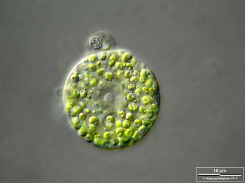
Scale bars indicate 50 µm (1), 25 µm (2, 4–6), 10 µm (3).
Six images. The coverslip print has disintegrated the sponge, making the amoeboid, totipotent cells visible. Totipotent means embryonic in the sense that each of these cells is capable of developing into a new sponge through successive mitotic divisions. Note that each cell has zoochlorellae!
Please click on < or > on the image edges or on the dots at the bottom edge of the images to browse through the slides!
Place name: Creek in Oder valley 100 km north east of Berlin (Germany)
Latitude: 53.135032 Longitude: 14.348738
Microscope Zeiss Universal, camera Olympus C7070WZ. DOF image.
© Wolfgang Bettighofer,
images under Creative Commons License V 3.0 (CC BY-NC-SA).
For permission to use of (high resolution) images please contact postmaster@protisten.de.
For further information about the image, please click here: Link to protisten.de page
Included On The Following Pages:
- Biota
- Eukaryota (eukaryotes)
- Unikonta
- Opisthokonta (opisthokonts)
- Metazoa (animals)
- Parazoa
- Porifera (sponges)
- Cellularia
- Demospongiae (demosponges)
- Haplosclerida
- Spongillina
- Spongillidae (freshwater sponges)
- Ephydatia
- Ephydatia fluviatilis
This image is not featured in any collections.
Source Information
- license
- cc-by-nc-sa-3.0
- copyright
- Wolfgang Bettighofer
- creator
- Wolfgang Bettighofer [email]
- original
- original media file
- visit source
- partner site
- protisten.de
- ID