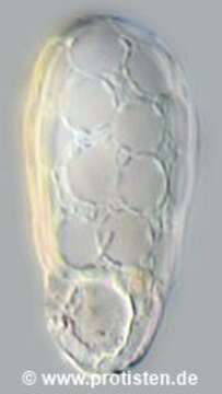
Image of Trinema Dujardin 1841
Description:
Sampling date 07/2018. Scale bars indicate 10 µm.
Two images.
First:Synoptic representation of the test (shell). Cell in vetral view.Second:Optical cross-section showing food vacuoles and the large nucleus in the fundus (posteriorly) with one nucleolus at position one o’clock.Please click on < or > on the image edges or on the dots at the bottom edge of the images to browse through the slides!
Place name: Lake Fuhlensee near Schilksee (Kiel, Germany)
Latitude: 54.43136338 Longitude: 10.16243935
Microscope Zeiss Axioplan, camera Olympus OM-D M5 MKII. DOF images.
© Wolfgang Bettighofer,
images under Creative Commons License V 3.0 (CC BY-NC-SA).
For permission to use of (high resolution) images please contact postmaster@protisten.de.
For further information about the image, please click here: Link to protisten.de page
Included On The Following Pages:
- Life
- Cellular
- Eukaryota
- SAR (Stramenopiles, Alveolates, Rhizaria)
- Rhizaria
- Cercozoa
- Imbricatea
- Silicofilosea
- Euglyphida
- Trinematidae
- Trinema
- Trinema enchelys
This image is not featured in any collections.
Source Information
- license
- cc-by-nc-sa-3.0
- copyright
- Wolfgang Bettighofer
- creator
- Wolfgang Bettighofer [email]
- original
- original media file
- visit source
- partner site
- protisten.de
- ID