Image of Trinema Dujardin 1841
Description:
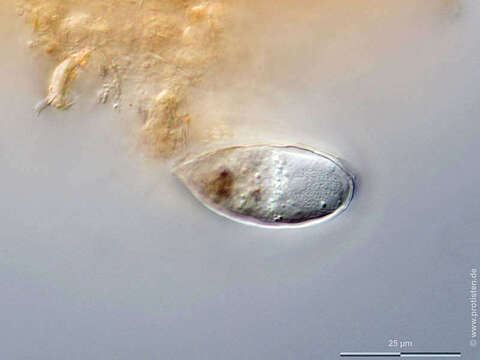
Sampling date 04/2021. Scale bars indicate 25 µm.
Three images.
First:Synoptic representation of the test (shell). Cell in lateral view.Second to fourth:Optical cross-sections, the last one shows the large nucleus with one nucleolus at position nine o’clock.Fouth:Region of the test´s neck with a large quartz grain, which is covered with silica produced by the amoeba.Fifth:A look inside the test shows that idiosomes are produced in reserve and stored in the cytoplasm in order to be used during cell division to build the test of the daughter cell.Please click on < or > on the image edges or on the dots at the bottom edge of the images to browse through the slides!
Place name: Pond near Großostheim (Germany)
Latitude: 49.88482168 Longitude: 9.09980822
Microscope Zeiss Axioplan, camera Olympus OM-D M5 MKII.
© Wolfgang Bettighofer,
images under Creative Commons License V 3.0 (CC BY-NC-SA).
For permission to use of (high resolution) images please contact postmaster@protisten.de.
For further information about the image, please click here: Link to protisten.de page
Included On The Following Pages:
- Life
- Cellular
- Eukaryota
- SAR (Stramenopiles, Alveolates, Rhizaria)
- Rhizaria
- Cercozoa
- Imbricatea
- Silicofilosea
- Euglyphida
- Trinematidae
- Trinema
- Trinema enchelys
This image is not featured in any collections.
Source Information
- license
- cc-by-nc-sa-3.0
- copyright
- Wolfgang Bettighofer
- creator
- Wolfgang Bettighofer [email]
- original
- original media file
- visit source
- partner site
- protisten.de
- ID