""
Description:
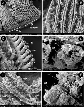
Figure 7A–F.Scanning electron micrographs of the ctenidium of Ochetoctena tomasi, A frontal face, frontal surface intact on the left, removed on the right to reveal tubules; B frontal surface with entrances to tubules arrowed; C single filament showing lateral and median tubules; D cross section of a single tubule; E long section of adjacent tubules; F bacterial bundles within bacteriocytes and paddle tipped filaments in the lumen of the tubule. bct bacteriocyte; spb spherical body; ptf paddle tipped filaments; fs frontal surface; fs (rem) frontal surface removed; lt lateral tubule; mt median tubule.
Included On The Following Pages:
- Life
- Cellular
- Eukaryota (eukaryotes)
- Opisthokonta (opisthokonts)
- Metazoa (animals)
- Bilateria
- Protostomia (protostomes)
- Spiralia (spiralians)
- Mollusca (molluscs)
- Bivalvia (mussels)
- Euheterodonta
- Imparidentia
- Lucinida (Lucinoida)
- Thyasiroidea
- Thyasiridae
- Ochetoctena
- Ochetoctena tomasi
This image is not featured in any collections.
Source Information
- license
- cc-by-3.0
- copyright
- P. Graham Oliver
- bibliographic citation
- Oliver P (2014) “TUBULAR GILLS” Extreme gill modification in the Thyasiroidea with the description of Ochetoctena tomasi gen. et sp. nov. (Bivalvia: Thyasiroidea) Zoosystematics and Evolution 90(2): 121–132
- original
- original media file
- visit source
- partner site
- Zoosystematics and Evolution
- ID