Image of Enterobacteriaceae
Description:
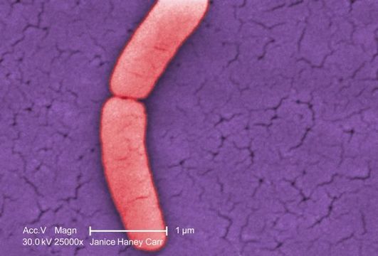
Under a very high magnification of 25000X, this colorized scanning electron micrograph (SEM) revealed the presence of a single Gram-negative Salmonella typhimurium bacterium, which was imaged right at the point where it was undergoing the process of cell division, resulting in the formation of two separate organisms. This dividing bacterium had been isolated from a pure culture. See PHIL 10994 for a black and white version of this image.
Created: 2009
Included On The Following Pages:
- Life (creatures)
- Cellular (cellular organisms)
- Bacteria
- Proteobacteria (Purple Bacteria & relatives)
- Gammaproteobacteria
- Enterobacterales
- Enterobacteriaceae
- Salmonella
This image is not featured in any collections.
Source Information
- license
- cc-publicdomain
- photographer
- Janice Haney Carr
- provider
- Public Health Image Library
- original
- original media file
- visit source
- partner site
- Public Health Image Library
- ID