Image of Dasymutilla Ashmead 1899
Description:
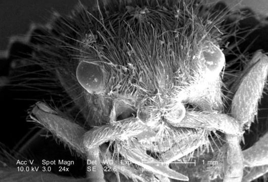
At a low magnification of only 24X, this scanning electron micrograph (SEM) showed the head region from an anterior view of a female velvet ant, Dasymutilla sp.. Note the two laterally positioned eyes, the anterior pair of antennae, each attached to the head by a rounded "scape", the numerous hairs or setae adorning almost all exterior surfaces, and the jointed legs, from which the insects Phylum Arthropoda is derived, i.e., Arthro = jointed, and poda leg. Also see PHIL 4638, 6363, and 6364 for photographs of the ant revealing its coloration, and velvety covering of external chitinous hairs
Created: 2007
Included On The Following Pages:
- Life (creatures)
- Cellular (cellular organisms)
- Eukaryota (eukaryotes)
- Opisthokonta (opisthokonts)
- Metazoa (Animal)
- Bilateria
- Protostomia (protostomes)
- Ecdysozoa (ecdysozoans)
- Arthropoda (arthropods)
- Pancrustacea
- Hexapoda (hexapods)
- Insecta (insects)
- Pterygota (winged insects)
- Neoptera (neopteran)
- Endopterygota (endopterygotes)
- Hymenoptera (wasps, bees, and ants)
- Apocrita (wasp)
- Aculeata
- Vespoidea (Yellowjackets and Hornets, Paper Wasps; Potter, Mason and Pollen Wasps and allies)
- Mutillidae (velvet ants)
- Dasymutilla
- Panarthropoda
This image is not featured in any collections.
Source Information
- license
- cc-publicdomain
- photographer
- Janice Carr
- provider
- Public Health Image Library
- original
- original media file
- visit source
- partner site
- Public Health Image Library
- ID