Image of Dasymutilla Ashmead 1899
Description:
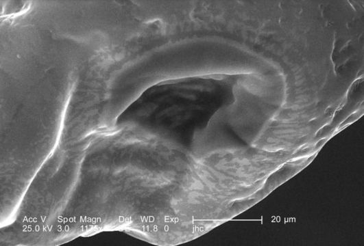
Under a high magnification of 1175X, this scanning electron micrograph (SEM) focused on the distal tip of a female velvet ants, Dasymutilla sp., stinger. In PHIL 9881 and 9882, lower magnifications places this image into a context, which allows you to appreciate its orientation. This is the stingers tip, which is encased in this bulbous sheath, which may act to protect the delicate sharp tip from being damaged, which would reduce its effectiveness when trying to penetrate its victim. Also see PHIL 4638, 6363, and 6364 for photographs of the ant revealing its coloration, and velvety covering of external chitinous hairs.
Created: 2007
Included On The Following Pages:
- Life (creatures)
- Cellular (cellular organisms)
- Eukaryota (eukaryotes)
- Opisthokonta (opisthokonts)
- Metazoa (Animal)
- Bilateria
- Protostomia (protostomes)
- Ecdysozoa (ecdysozoans)
- Arthropoda (arthropods)
- Pancrustacea
- Hexapoda (hexapods)
- Insecta (insects)
- Pterygota (winged insects)
- Neoptera (neopteran)
- Endopterygota (endopterygotes)
- Hymenoptera (wasps, bees, and ants)
- Apocrita (wasp)
- Aculeata
- Vespoidea (Yellowjackets and Hornets, Paper Wasps; Potter, Mason and Pollen Wasps and allies)
- Mutillidae (velvet ants)
- Dasymutilla
- Panarthropoda
This image is not featured in any collections.
Source Information
- license
- cc-publicdomain
- photographer
- Janice Carr
- provider
- Public Health Image Library
- original
- original media file
- visit source
- partner site
- Public Health Image Library
- ID