Image of human body louse
Description:
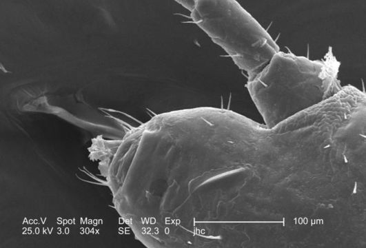
Under a moderately low magnification of 304x, this 2006 scanning electron micrograph (SEM) depicted an enlarged inferior view of the mouthparts found on the cephalic, or head region of a male body louse, Pediculus humanus var. corporis. The floral-like structure in this field of view represents the labial funnel, or haustellum, which is a tube-like structure, formed from what is believed to be a modification of the labium, but is armed with teeth, which act to grab, and hold on to the skin surface of its host while obtaining its blood meal. The insect pierces the hosts skin with its sharply-pointed set of three stylets, and together, form what is termed the fascicle, which is hidden from view in this image. Note PHIL# 9214, 9216, in which case a louse is in its feeding mode on the skin surface of its host. Also note the scape and pedicle, or the respective proximal segments of the insect's left antenna in this view as well.
Created: 2006
Included On The Following Pages:
- Life (creatures)
- Cellular (cellular organisms)
- Eukaryota (eukaryotes)
- Opisthokonta (opisthokonts)
- Metazoa (Animal)
- Bilateria
- Protostomia (protostomes)
- Ecdysozoa (ecdysozoans)
- Arthropoda (arthropods)
- Pancrustacea
- Hexapoda (hexapods)
- Insecta (insects)
- Pterygota (winged insects)
- Neoptera (neopteran)
- Paraneoptera
- Psocodea (bark lice, book lice and true lice)
- Troctomorpha (book louse)
- Pediculidae (primate body lice)
- Pediculus
- Pediculus humanus (human body louse)
- Panarthropoda
- Nanopsocetae
This image is not featured in any collections.
Source Information
- license
- cc-publicdomain
- photographer
- Janice Carr
- provider
- Public Health Image Library
- original
- original media file
- visit source
- partner site
- Public Health Image Library
- ID