Image of Yersinia
Description:
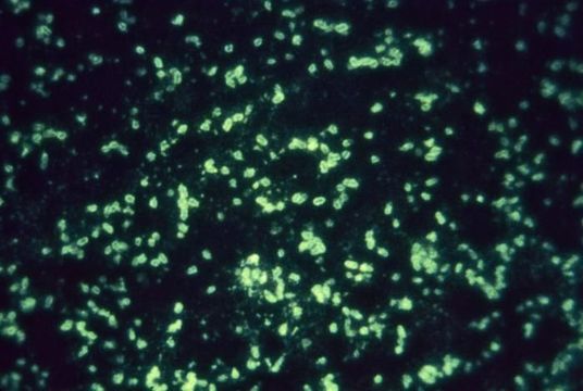
This micrograph was stained using a fluorescent antibody staining technique (FA), which uses the specific conjugated antiserum to Fraction 1 (F1) antigen of Yersinia pestis to identify the antigens present in animal tissues, and appropriate cultures.
Created: 1993
Included On The Following Pages:
- Life (creatures)
- Cellular (cellular organisms)
- Bacteria
- Proteobacteria (Purple Bacteria & relatives)
- Gammaproteobacteria
- Enterobacterales
- Yersinia
- Yersinia pestis
- Enterobacteriaceae
This image is not featured in any collections.
Source Information
- license
- cc-publicdomain
- provider
- Public Health Image Library
- original
- original media file
- visit source
- partner site
- Public Health Image Library
- ID