Image of Entamoebidae
Description:
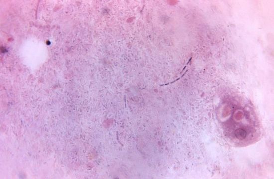
Magnified 675X, this photomicrograph revealed the presence of a parasitic Entamoeba histolytica trophozoite, which contained vacuolated cytoplasm, within which were two red blood cells (RBCs), and a pyknotic body. Entamoeba histolytica/Entamoeba dispar trophozoites have a single nucleus, which have a centrally placed karyosome and uniformly distributed peripheral chromatin. This typical appearance of the nucleus is not always observed as some trophozoites can have nuclei with an eccentric karyosome and unevenly distributed peripheral chromatin. The cytoplasm has a granular or "ground-glass" appearance. E. histolytica/E. dispar trophozoites usually measure 15µm - 20µm (range 10µm - 60µm), tending to be more elongated in diarrheal stool.
Created: 1971
Included On The Following Pages:
- Life (biota)
- Cellular
- Eukaryota (eukaryotes)
- Amoebozoa (amoeboid protists)
- Archamoebae
- Entamoebidae
- Entamoeba
- Entamoeba histolytica (Amoebic Dysentery)
This image is not featured in any collections.
Source Information
- license
- cc-publicdomain
- provider
- Public Health Image Library
- original
- original media file
- visit source
- partner site
- Public Health Image Library
- ID