Image of Petricola Lamarck 1801
Description:
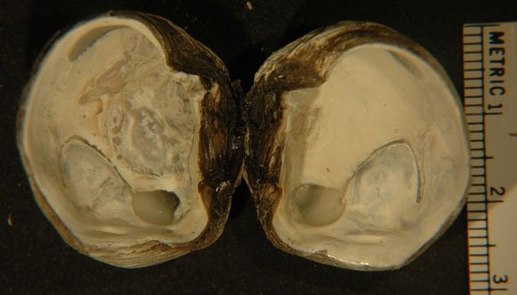
In this inside view, the anterior end of the shell is up and posterior is down. The well-defined pallial line is visible in both shells, as well as the deep pallial sinus near the posterior end. Both the anterior and posterior adductor muscle scars are visible near the hinge, with the posterior scar most evident. A good deal of brown, adhering periostracum can still be seen near the hinge of this individual.
Included On The Following Pages:
- Life
- Cellular
- Eukaryota (eukaryotes)
- Opisthokonta (opisthokonts)
- Metazoa (animals)
- Bilateria
- Protostomia (protostomes)
- Spiralia (spiralians)
- Mollusca (molluscs)
- Bivalvia (mussels)
- Euheterodonta
- Imparidentia
- Venerida
- Veneroidea
- Veneridae (venus clams)
- Petricolinae
- Petricola
- Petricola carditoides
- Heterodonta
This image is not featured in any collections.
Source Information
- license
- cc-by-nc-sa
- copyright
- Rosario Beach Marine Laboratory
- provider
- Invertebrates of the Salish Sea
- original
- original media file
- visit source
- partner site
- Invertebrates of the Salish Sea
- ID