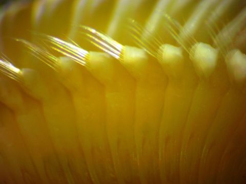
Image of Paralvinella palmiformis Desbruyères & Laubier 1986
Description:
This closeup of the parapodia on a posterior section of the body shows the well-developed notopodium (dorsal portion) with well-developed notosetae. The ventral portion of the parapodium (neuropodium, top of photo) appears to be simply a flattened ridge with no neurosetae visible such as was seen on the anterior portion, although the neuropodial ridge is better developed. The species description states that neurosetae are present on the posterior portion of the body so they should be visible here. However, in Terebellids, which are other members of this Order, the neurosetae are actually very short uncini which are hard to see. That may be the case here as well.
Included On The Following Pages:
- Life (creatures)
- Cellular (cellular organisms)
- Eukaryota (eukaryotes)
- Opisthokonta (opisthokonts)
- Metazoa (Animal)
- Bilateria
- Protostomia (protostomes)
- Spiralia (spiralians)
- Annelida (segmented worms)
- Sedentaria
- Terebelliformia
- Alvinellidae (Pompeii worms)
- Paralvinella
- Paralvinella palmiformis
This image is not featured in any collections.
Source Information
- license
- cc-by-nc-sa
- copyright
- Rosario Beach Marine Laboratory
- provider
- Invertebrates of the Salish Sea
- original
- original media file
- visit source
- partner site
- Invertebrates of the Salish Sea
- ID