Stubby microvilli (mv) arising from a primary oocyte (00) in the lumen of the ovary. The apical surface of the ovarian...
Description:
Stubby microvilli (mv) arising from a primary oocyte (00) in the lumen of the ovary. The apical surface of the ovarian...
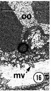
Stubby microvilli (mv) arising from a primary oocyte (00) in the lumen of the ovary. The apical surface of the ovarian epithelium is depicted at the bottom of the micrograph. Note that the oocyte lacks well developed extracellular coats or surrounding follicle cells. Scale bar, 1 urn.
Included On The Following Pages:
- Life (creatures)
- Cellular (cellular organisms)
- Eukaryota (eukaryotes)
- Opisthokonta (opisthokonts)
- Metazoa (Animal)
- Bilateria
- Protostomia (protostomes)
- Spiralia (spiralians)
- Nemertea (ribbon worms)
- Hoplonemertea
- Monostilifera
- Carcinonemertidae
- Carcinonemertes
- Carcinonemertes epialti
- Enopla
- Eumonostilifera
This image is not featured in any collections.
Source Information
- license
- cc-by-nc
- copyright
- Stricker, 1986
- photographer
- Stricker
- publisher
- Keen, Eric
- provider
- Nemertea
- original
- original media file
- visit source
- partner site
- Nemertea
- ID