Transmission electron micrograph (TEM) of three small germinal cells (ge) in the subepidermal region of the ovarian epithelium..
Description:
Transmission electron micrograph (TEM) of three small germinal cells (ge) in the subepidermal region of the ovarian epithelium..
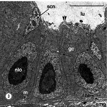
Transmission electron micrograph (TEM) of three small germinal cells (ge) in the subepidermal region of the ovarian epithelium. The double arrowheads mark apical processes of somatic cells that cover the germinal cells. hi, basal lamina; iu, lumen of ovary; m, myofilaments; nlo, nucleolar body; scn, somatic cell nucleus; smg, submuscular glands. Scale bar, 5 micrometers.
Included On The Following Pages:
- Life (creatures)
- Cellular (cellular organisms)
- Eukaryota (eukaryotes)
- Opisthokonta (opisthokonts)
- Metazoa (Animal)
- Bilateria
- Protostomia (protostomes)
- Spiralia (spiralians)
- Nemertea (ribbon worms)
- Hoplonemertea
- Monostilifera
- Carcinonemertidae
- Carcinonemertes
- Carcinonemertes epialti
- Enopla
- Eumonostilifera
This image is not featured in any collections.
Source Information
- license
- cc-by-nc
- copyright
- Stricker, 1986
- photographer
- Stricker
- publisher
- Keen, Eric
- provider
- Nemertea
- original
- original media file
- visit source
- partner site
- Nemertea
- ID