Plate 5
Description:
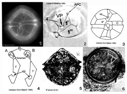
Plate 5. Alexandrium ostenfeldii. Figs. 1-3. LM. Fig. 1. Ventral view. Cell large and nearly spherical. Cingulum deeply excavated. Epitheca broad and convex-conical. Hypotheca hemispherical with an obliquely flattened antapex. Fig. 2. Epitheca: apical view. Ventral pore (vp) large and distinct. First apical plate (1') forms a 90 degree angle at the point where vp and 4' plate come in contact. Apical pore complex (APC) with comma-shaped foramen. Figs. 3-4. Line drawings. Fig. 3. Ventral view: 6'' plate wider than high. Cingulum (C) slightly excavated. Fig. 4. APC and 1' plate: a. Po in direct contact with 1'; b. Po in indirect contact with 1' via thin suture. Fig. 5. LM: vegetative cell. Small equatorial nucleus (n). Fig. 6. LM: temporary cyst large and spherical, covered in mucilage. Nucleus visible (arrowhead)(Mackenzie et al. 1996).
Included On The Following Pages:
- Life (creatures)
- Cellular (cellular organisms)
- Eukaryota (eukaryotes)
- SAR (Stramenopiles, Alveolates, Rhizaria)
- Alveolata (alveolates)
- Dinophyceae
- Gonyaulacales
- Gonyaulacaceae
- Alexandrium
- Alexandrium ostenfeldii
- Dinoflagellata (dinoflagellates)
This image is not featured in any collections.
Source Information
- license
- cc-publicdomain
- bibliographic citation
- Faust, Maria A. and Rose A. Gulledge. Identifying Harmful Marine Dinoflagellates. Smithsonian Contributions from the United States National Herbarium, volume 42: 1-144 (including 48 plates, 1 figure and 1 table).
- original
- original media file
- visit source
- partner site
- NMNH Marine Dinoflagellates
- ID