Plate 12
Description:
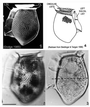
Plate 12. Dinophysis acuta. Fig. 1. SEM: lateral view. Cell oblong and robust; theca heavily areolated. Well developed cingular lists (CL) and left sulcal list (LSL). Pointed antapex. Figs. 2-3. LM: lateral view (from Larsen & Moestrup 1992: fig.s 2a,d; scale bars=20 _). Fig. 2. Large areolae, each with a pore (arrows). Fig. 3. Widest point below mid-section (dashed line) aligned with third sulcal rib (arrow). Fig. 4. Line drawing.
Included On The Following Pages:
- Life
- Cellular
- Eukaryota (eukaryotes)
- SAR (Stramenopiles, Alveolates, Rhizaria)
- Alveolata (alveolates)
- Dinophyceae
- Dinophysiales
- Dinophysiaceae
- Dinophysis
- Dinophysis acuta
- Dinoflagellata (dinoflagellates)
This image is not featured in any collections.
Source Information
- license
- cc-publicdomain
- bibliographic citation
- Faust, Maria A. and Rose A. Gulledge. Identifying Harmful Marine Dinoflagellates. Smithsonian Contributions from the United States National Herbarium, volume 42: 1-144 (including 48 plates, 1 figure and 1 table).
- original
- original media file
- visit source
- partner site
- NMNH Marine Dinoflagellates
- ID